


We commonly have patients and/or parents question why we obtain a panorex as part of our comprehensive assessment for oral surgical procedures. A panorex is an x–ray that provides a full view of the upper and lower jaws, teeth, temporomandibular joints (TMJs) and sinuses. Without this critical piece of data, we can sometimes miss pathologic lesion, such as tumors or cysts, extra teeth, and a whole host of other conditions that need surgical treatment in a timely fashion. Just having your typical “PA” x-rays that a general dentist obtains is insufficient for our purposes – this is clearly demonstrated below.
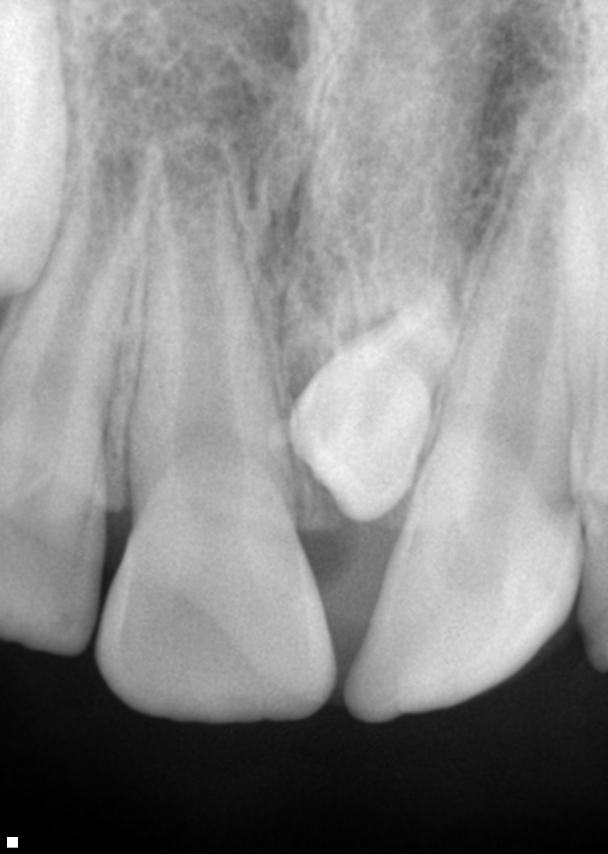
Below you see a “PA”, that is a much smaller xray designed to only look at teeth and not the entire face.
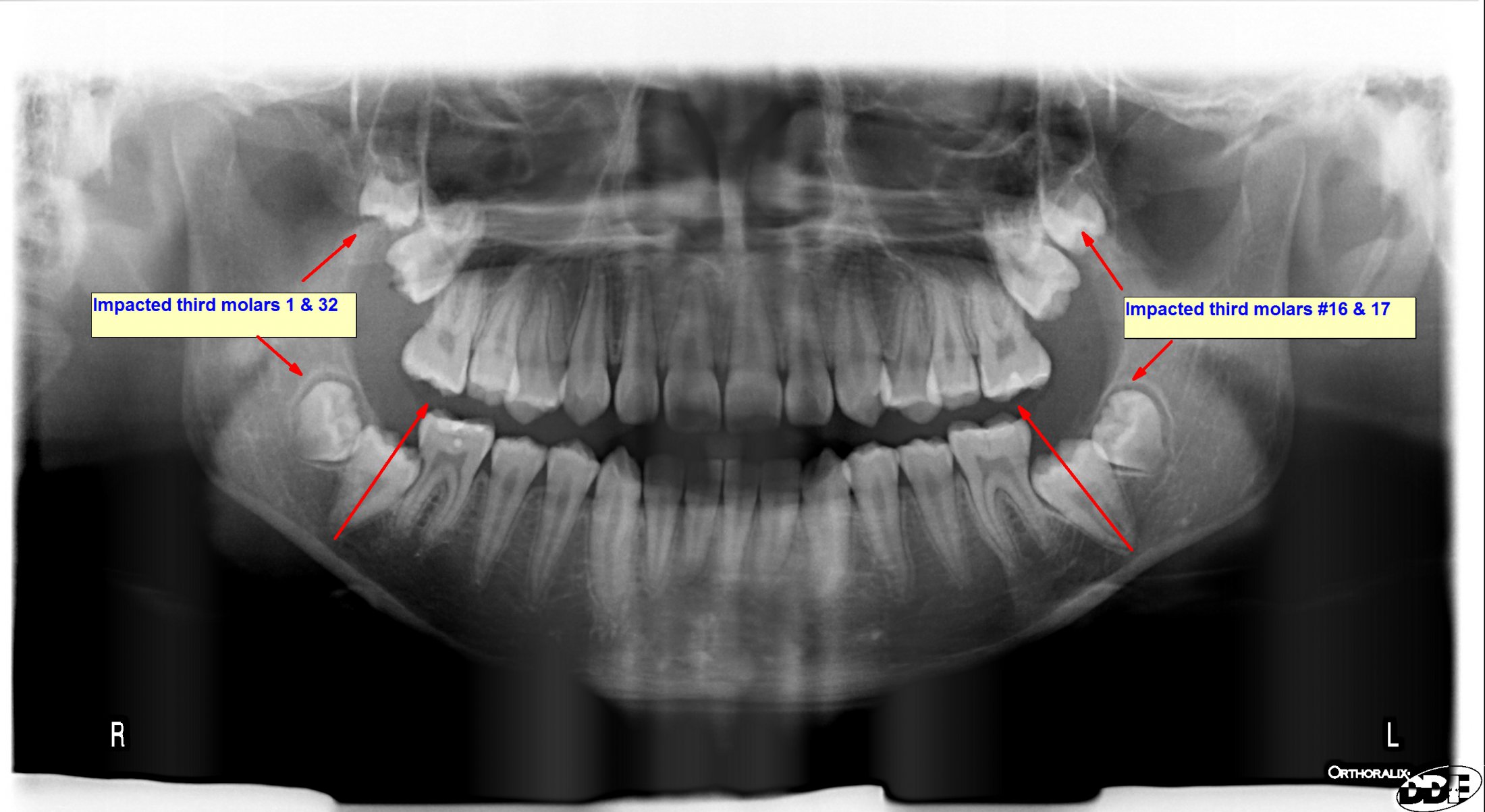
A “panorex” as seen below, is a larger xray designed to see more than teeth.
The case below is a perfect example as to why we obtain a panorex in the early teenage years to rule out pathology and other abnormalities – such as disturbances in tooth eruption.
Today we operated on a 13 year old that presented to their pedodontist for what they thought would be a routine follow-up. The parents did not even realize he was missing a bunch of his back teeth? As a part of the routine consultation a panorex was taken by the pedodontist and to their surprise they noted that he had 8 impacted teeth: four impacted 2nd molars, that were impacted due to his four impacted 3rd molars! So what did we do? He was referred to us for the extraction of his third molars in order to create space for the impacted 2nd molars. Additionally, when we removed the impacted third molars, we surgically can “unlock” or partially reposition the impacted 2nd molars into a more ideal position….with the hope of having them erupt spontaneously – (without the need for orthodontics.)
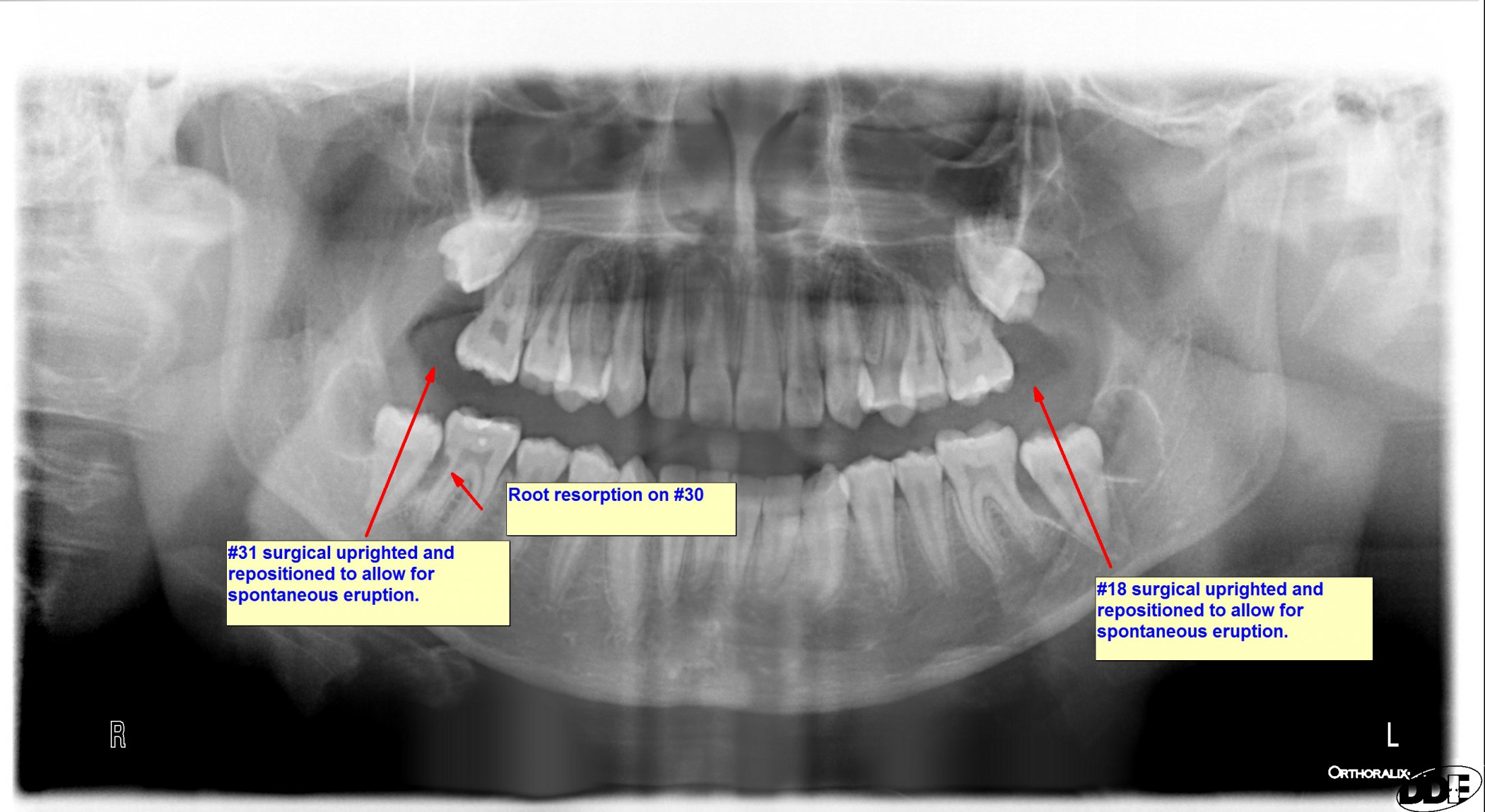
So the patient underwent the extraction of all third molars 1, 16. 17 & 32; with surgical uprighting of second molars #18 & 31. The RED ARROW shows the axis of the lower 2nd molars 18 & 31. With careful attention you can see the axis has changed from the pre-to the post-operative panorex that was taken immediately after surgery.
Additionally you may notice a notch on the back side of the lower right 1st molars #30 (as we refer to the distal aspect of root #30)….This is “root resorption” or damage that was being caused by the impacted 2nd molar #31. Now that we have “unlocked” or uprighted #31, hopefully the damage will subside and the patient will not need a root canal on #30 when he grows up.
This is why panorex xrays are so critical in early diagnosis of oral pathologies before there is permanent damage to someone oral health. Timing is everything here. We are hoping by treating the patient at age 13, we can prevent any future damage that may lead to premature of loss of his adult teeth. The surgical uprighting of the #18 & 31 may also prevent him from having to get orthodontic treatment (braces) – saving him and his family thousands in unnecessary expense.
